Clearing the confusion
04 April 2013
Volume 29 · Issue 4

Colin Campbell advocates the use of cone beam computed tomography.

As an implant surgeon I use a considerable amount of CBCT within my field of expertise. My practice uses CBCT in the majority of cases for implant planning and increasingly for other disciplines such as endodontics, orthodontics, oral surgery, implant surgery and periodontics. The practice also takes referrals from the local hospital, which doesn’t have a CBCT machine for its maxillofacial unit and orthodontics.

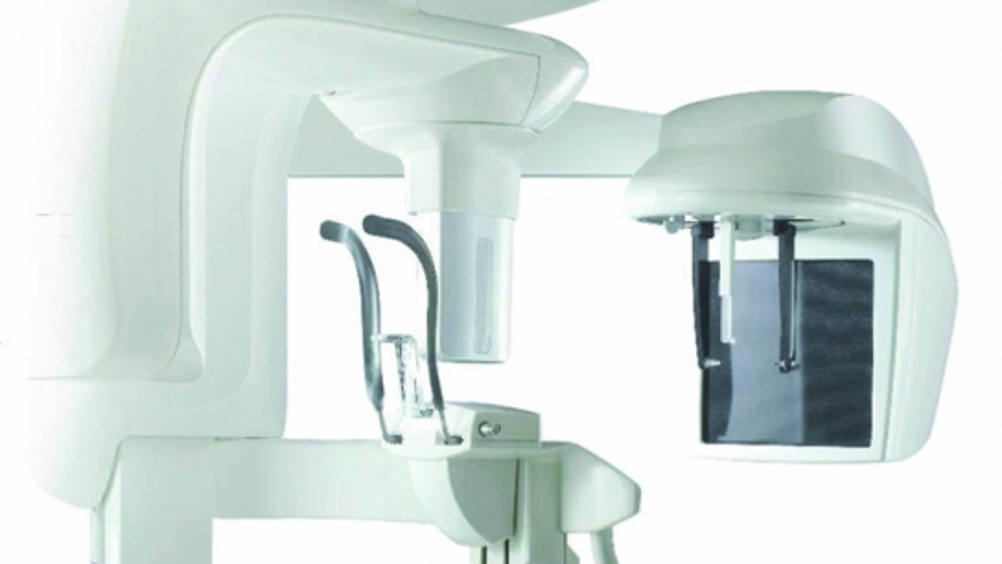
Cone beam computer tomography (CBCT) is an X-ray modality, a radiography examination for patients which provides a considerable amount of detail as well as significant advantages over traditional two dimensional methods. I have used the CS 9000 3D system from Carestream Dental for the last three years.

Cone beam CT is a radiography modality that was specifically designed for examination of the head and neck area. It differs from medical grade CT in two main ways. First, patients are seated or standing when it is taken (similar to a digital panoramic radiograph) as opposed to lying down; and second, CBCT has a dramatically reduced dose.

The image quality of CBCT can be impressive, though of course it does depend on the system being used. The quality of image increases with voxel (volumetric pixel, which is the 3D equivalent of a pixel) size; the smaller the voxel, the greater the results. With the CS 9000 3D system, the voxel size is extremely small, approximately 0.07mm, ensuring a very detailed image. Scanning is also very quick and efficient - a single scan takes just 30 seconds, and a unit is easy to use, allowing the task to be delegated to team members at the practice. In fact, in three years and hundreds of CBCTs, we have never had to repeat a CBCT because of substandard image quality.

The image is created from a data file and pushed through a software converter almost on a one-to-one ratio. The benefits of 3D over traditional imaging are many and varied. In particular, there is huge scope for manipulation of the images. They can be turned, flipped, enhanced, or modified, or implant stents can be added.

I think there is still confusion amongst some people who don’t use Cone Beam CT, whereby they think it is on a par with medical grade CT scans. Two radiation dose comparison studies (IRSN and Ludlow) have shown the reduced risk of focused field 3D imaging (focused field effectively meaning ‘a smaller field of view’) using CBCT.

For a standard digital panoramic examination, the effective dose is estimated to be 7-22µSv, which is equivalent to approximately one to three days of radiation. A 3D exam performed with the CS 9000 3D system has the same radiation equivalent, because the effective dose is around 5-19µSv. The variation in dose depends on where the field of view is positioned. For example, the posterior mandible would be more radiographically sensitive than the premaxilla. Some other 3D systems are effective between 68-600µSv. There is a huge variation with the different systems, which is something that needs to be taken into account prior to making a purchase.

The introduction of focused field and 3D imaging is significant in terms of the improvements to patient safety and diagnostic capability. Prior to these systems, it was traditional to scan a full arch or even upper and lower arches simultaneously, with no possibility for adjustment of the size of the area scanned. This led to concern about overdosing patients because they were being examined in areas not required. In the past few years, developments have been made in focused field of view imaging systems that allow clinicians to concentrate on smaller areas of the patient specific to the diagnosis.

There are CBCT systems that can focus on a field of view of 50mm x 37mm, which allows the examination of just over a sextant. Full arches can still be achieved, however, the unit simply scans the patient three times at a lower dose and stitches them together to produce an image that can be used as a conventional full arch.

A small focus field of view reduces the radiation exposure and provides a higher resolution image for more precise visualisation of the dental anatomy. This opens up useage for many indications while confining exposure directly to the area of interest, reducing patient dose and aligning with the ALRA (as low as reasonably achieveable) principle.

Looking at CBCT in dentistry, both from a dosage and risk perspective of the patient and for functionality in the practice, I believe it truly transforms practice life and the quality of dentistry you can offer.