Increasing precision
24 May 2022

While a poor craftsman blames his tools, there are certainly gains to be made by understanding and incorporating technological innovations into our repertoire.  

Increasing accuracy and precision is an evergreen goal among dental practitioners. From more precise diagnostics to more precise treatment, and even how we communicate with patients and colleagues – precision can be said to influence everything we do. While a poor craftsman blames his tools, there are certainly gains to be made by understanding and incorporating technological innovations into our repertoire.  

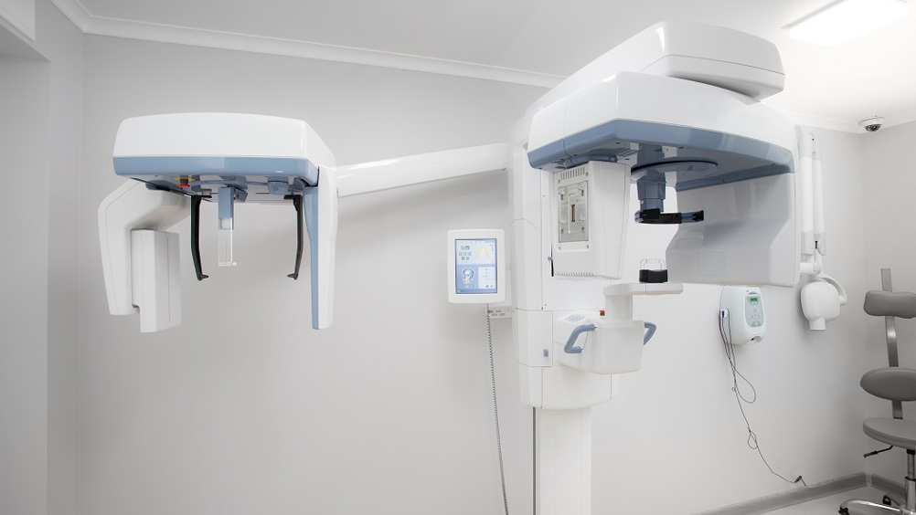
An increasingly popular and widespread example is the use of cone beam computed tomography (CBCT) in conjunction with intra-oral optical scanning (IOS). In a typical digital dental workflow, CBCT is used to construct 3D rendering of a patient’s oral cavity and surrounding tissue. Compared to the traditional two-dimensional CT scan, three-dimensional data is transformative in facilitating computer aided design and manufacture (CAD/CAM) with wide ranging applications in dentistry, from design and manufacture of full arch bridges and guided surgery stents to construction of prostheses and even 3D printed biomaterials. Anatomical features that can be masked or obscured in a flat image are much more readily identifiable in 3D. For example, the root structure of teeth is more easily visible in CBCT imaging, which can make detecting apical anomalies or lesions more achievable.[1] This has obvious implications for surgical planning and the avoidance of complications, as complex features that may have been easily missed can now be mapped, measured, and accounted for prior to any invasive procedure. Quite simply, this information gives dentists a chance to change or adapt their approach in advance, for optimal outcomes. For example, 3D printed surgical guides are transforming the accuracy and precision of implant placements whilst reducing the risk of collateral damage to surrounding vital structures.

CBCT scans still require an experienced clinician to interpret. Common dental implant procedures necessitate that vital anatomical structures, such as the neurovascular bundle, are located and protected with accuracy. One widely used way to achieve this is for clinicians to label the inferior alveolar canal in cross-sectional slices using 3D imaging software to produce segmentation of the canal – a process that is labour-intensive and time-consuming.[2] Using CBCT scans, efforts are currently underway to train artificial intelligences to carry out mandibular canal segmentation automatically, which if successful, could reduce workloads and increase efficiency. Teams currently developing deep learning algorithms to achieve this are reporting promising results, and this may prove to be but the tip of the iceberg in what deep learning technologies can bring to dentistry.2, [3] Of course, as with any application of algorithmic automation, a note of caution is warranted, as among other things it is all too easy to bias the dataset used to train the algorithm as recent news stories will attest.

Driven by the development of existing and new underlying technologies, digital dentistry is constantly evolving. Sometimes this occurs in unexpected directions, such as adapting oral scans into NFTs (non-fungible tokens). William Shatner, of Captain Kirk fame, infamously converted x-rays of his teeth into an NFT.[4] At least one enterprising dentist has used intraoral scans as the basis for NFT art, whilst elsewhere using dental-inspired NFT art to partially fund a dental charity campaign has been attempted.[5], [6] Whether such early adoption of a controversial, headline-generating technology will ultimately prove successful is hard to say, but it does illustrate that burgeoning technologies create opportunities for entrepreneurialism.

Recognising the potential of new technologies and methodologies is merely the first step to utilising and fully exploiting the advantages they present. Dentistry is fundamentally somewhat conservative, and for good reason; innovations have to prove not only their utility but also their safety and reliability. A careful balance must be struck between embracing new ways of working, without getting ahead of the evidence. Research papers are helpful in getting a scientific insight into approaches, and as data accumulates, we get a clearer understanding of what is and is not working and why (sometimes with surprising upsets to established wisdom). However, keeping abreast of research can be time consuming (let alone evaluating it), and papers often do not provide definitive solutions or answer certain questions a clinician may have. Looking to experienced clinicians who have remained on the cutting-edge is a wise choice, if you want to embrace the latest and greatest methods with improved certainty.

Ucer Education, has been at the forefront of postgraduate education in UK implantology for over a quarter century. Led by specialist oral surgeon, Professor Cemal Ucer, Ucer Education’s Postgraduate Certificate (PGCert) in Implant Dentistry (EduQual Level 7) is an outstanding opportunity for clinicians looking to advance their skills and knowledge. The course lays down a solid grounding in technical competencies and theory. Moreover, you will gain a current understanding in the latest and greatest techniques and technologies including different implant systems and their provision, full digital workflow including CBCT scans, 3D treatment planning and printing, and much more. The highly knowledgeable team have the experience to answer almost any question you might have, and provide real world insight into all aspects of implantology.

Technology is a tool, and as with any tool it must be wielded with knowledge and skill to achieve the best results. Understanding when to use a particular tool and how to get the most out of a given approach – for that there are no quick and easy technological shortcuts – it takes learning and experience, and the irreplaceable human element of good judgement.

[1] Kaasalainen T., Ekholm M., Siiskonen T., Kortesniemi M. Dental cone beam CT: an updated review. Physica Medica. 2021; 88: 193-217. https://doi.org/10.1016/j.ejmp.2021.07.007 February 12, 2022.

[2] Jaskari J., Sahlsten J., Järnstedt J., Mehtonen H., Karhu K., Sundqvist S., Hietanen A., Varjonen V., Mattila V., Kaski K. Deep learning method for mandibular canal segmentation in dental cone beam computed tomography volumes. Scientific Reports. 2020; 10: 5842. https://doi.org/10.1038/s41598-020-62321-3 February 12, 2022.

[3] Lahoud P., Diels S., Niclaes L., Van Aelst S., Willems H., Van Gerven A., Quirynen M., Jacobs R. Developmment and validation of a novel artificial intelligence driven tool for accurate mandibular canal segmentation on CBCT. Journal of Dentistry. 2022; 116: 103891. https://doi.org/10.1016/j.jdent.2021.103891 February 12, 2022.

[4] Shatner W [@WilliamShatner]. Yes.  You want a pack of my NFT trading cards? I put X-ray images of my tooth from 1953 up as an NFT. Who does something like that? Me! [Tweet]. Twitter. February 20, 2021. https://twitter.com/WilliamShatner/status/1363162833932886017?s=20&t=mf2tHLlE-Z3Q-bOzIRc6zg February 11, 2022.

[5] The NFTeeth Project. http://nfteeth.info/The-NFTeeth-Project/ February 11, 2022.

[6] Dentacoin. NFTs by dentacoin: supporting dental charity causes. https://dentacoin.medium.com/nfts-by-dentacoin-supporting-dental-charity-causes-64f92b66279c February 11, 2022.