
Giuliano Fragola presents a complex premolar root fracture case using the new CS 8200 3D Neo Edition CBCT system from Carestream Dental.
A 45-year-old female patient presented with pain in the teeth of the first quadrant and the sensation of slight mobility when she chewed. Her overall condition was good – she was a non-smoker, with good oral hygiene. The dental history revealed that she had undergone endodontic treatment about 15 years previously, and she had a full contour crown in place, which was causing no issues.
In trying to identify the source of the pain, extensive visual inspection and percussions were carried out, but these alone were not enough to diagnose the source of the pain. A 3D x-ray was therefore indicated to assess potential subgingival or bone complications. The new CS 8200 3D Neo Edition from Carestream Dental was used to take the image, with the MAR tool (Metal Artefact Removal) delivering precise and very clear images for diagnosis and treatment planning. It can be very difficult to see a root fracture of this nature due to reflections from existing crowns, which are typically placed over endodontically-treated teeth, but this technology overcomes this problem. Having used digital technologies from Carestream Dental since the late 1990s, I completely trust the quality of the equipment available.
It was also possible to narrow the FOV to only the area in question and the lateral scout view played an important role in enabling us to check the targeting of the desired area, avoiding the risk of retakes. The low dose mode was used to minimise exposure while still delivering very good image quality.
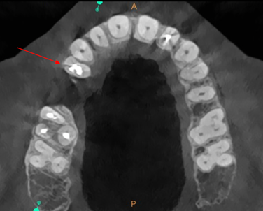
Figure 1a - Axial view of the maxilla, showing the root fracture of tooth 14 (red arrow)
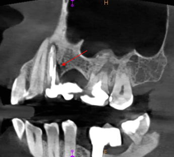
Figure 1b – Sagittal view showing the root fracture of tooth 14, the bone loss associated (red arrow) and an apical lesion
The images demonstrated bone loss around the sites of the 14 and 16 [Figures 1a and 1b], which explained the mobility and positive percussion. Using different 3D views, a complex root fracture was diagnosed in the first premolar (14) [Figure 2], along with complete bone loss between the roots of the first maxillary molar (16) and a small sinus floor bone perforation [Figure 3]. This provides a very useful tool for the clinician, facilitating communication with the patient and encouraging their engagement with treatment from the very beginning. With CS 8200 3D Neo Edition image, it was easy for the patient to visualise the problem and understand the proposed treatment, so she felt confident to accept it and proceed straightaway.
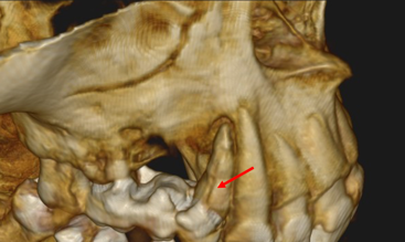
Figure 2 -3D rendering showing the vertical root fracture on the vestibular side of tooth 14 (red arrow)
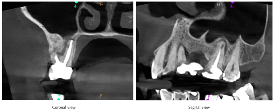 Figure 3 - Coronal and sagittal views of tooth 16, showing the dentine and bone resorption on the palatal root and the sinus floor perforation
There was only one viable treatment option available in this case – the 14 and 16 both had a hopeless prognosis and so would require extraction and restoration. The procedure was explained to the patient in full, including benefits, limitations and potential risks to ensure informed consent to proceed.
Both teeth were extracted as atraumatically as possible and then left to heal for approximately six months, when dental implants would be placed. The patient refused a removable denture, preferring restoration with an implant-retained prosthesis instead. Given the complexity of this case, a new CBCT scan was indicated post hard and soft tissue healing to assess bone volume remaining for implant placement planning.
The patient was very satisfied with the outcome of treatment, appreciating the quick and precise diagnosis and treatment delivery based on the information obtained. She commented, “I’m absolutely impressed by the images shown by the dentist with the fracture lines. I completely understand that extraction is the only option.”
At this stage, we are waiting for the soft and hard tissue to fully heal before deciding how to proceed. A new CBCT scan may be indicated post healing to analyse the bone volume and density, and to support the planning procedure for implant placement and restoration. This scan would be used to help determine whether bone grafting will be required and if it can be delivered simultaneously with implant placement. Even with the second CBCT scan, the patient would still be exposed to less radiation in the long-term compared to the alternative – multiple periapical x-rays at different angles. The CBCT low dose mode is ideal for reducing exposure without compromising image quality.
From a professional perspective, good results were achieved in this case. The new CBCT scanner was instrumental in ensuring accuracy and confidence of every stage in the treatment, from diagnosis to treatment planning and delivery. It was also beneficial in supporting and improving communication with the patient, enhancing their understanding of the procedure and ensuring quick but confident informed consent. The CS 8200 3D Neo Edition helps to obtain precise and high-quality images with the lowest possible dose of radiation. Being able to select the FOV with a simple and quick view shot, and to manage the scattering with digital formatting are further benefits.
For more information, contact Carestream Dental on 0800 169 9692 or visit www.carestreamdental.co.uk